Summer School 2025

Advanced Plant Imaging – API : From super-resolution to fluorescence lifetime imaging microscopy

June 29 - July 4, 2025 – Versailles (France)

Saclay Plant Sciences (SPS)

This Summer school is organized by the Saclay Plant Sciences (SPS) network, one of the largest European plant sciences communities and will be hosted by the cytology and imaging platform of the Institut Jean Pierre Bourgin - Plant Sciences in Versailles.

Microscopy is a fundamental tool for understanding the functioning of plants at the cellular to molecular scale. Recent technological advances (super-resolution, fluorescence lifetime imaging, biosensors...) now make it possible to address new scientific questions. This Summer School will provide theoretical and practical insights into these aspects for 20 outstanding and enthusiastic PhD students or young postdoctoral researchers divided in small groups. Participants will have the opportunity to discuss the advantages and disadvantages of different modalities, sample preparation, image acquisition, and data processing throughout the week.

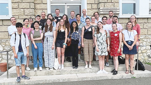
Summer School 2024 - Photo de groupe
Summer School 2024

Provisional program and speakers

The Summer School will include:

Cross-section of pomegranate stem labeled with propidium iodide. Observation with a confocal laser scanning microscope. © Romain Le Bars @ Imagerie-Gif

> A set of theoretical lectures (~9 hours) on the major issues in advanced imaging: nanoscopy (super resolution), fluorescence lifetime analysis, ratiometry analysis, their uses, advantages or limitations, data processing, etc. The lectures will be given by experts in the field, who will be available for discussions during the Summer School, giving the participants an insight into the latest research findings and identifying key open questions in the field.

Confirmed speakers:
Joachim Goedhart (University of Amsterdam, The Netherlands)
Julien Gronnier (Technical University of Munich, Germany)
Kalina Haas (Jean-Pierre Bourgin Institute - Plant Sciences, Versailles, France)
Eric Hosy (Interdisciplinary Institute for NeuroScience, Bordeaux, France)
Joris Sprakel (Wageningen University & Research, The Netherlands)

> A set of practical sessions (~24 hours + Restitution) to tackle real-life approaches on plant samples and compare different modalities of conventional confocal microscopy, super-resolution and fluorescence lifetime analysis. All Summer School participants will have access to all practical sessions, in groups of 3 or 4, with one (or more) expert per system. Given the advanced technologies covered, the Summer School is aimed at PhD students or young post-docs who already have practical experience in confocal microscopy.

Topics will include:

Observation of the synaptonemal complex structure during meiosis using confocal (left) and STED microscopy (right) © Aurélie Hurel / Mathilde Grelon

1) Lifetime analysis (FLIM) and super-resolved STED microscopy on a LEICA stellaris 8, excitation by white laser, with a STED section consisting of a depletion laser at 775nm, Tau STED and Gated STED modes and 3D STED modality. Tau and Gated STED can be used with the Tau Sense tool or with FALCON. This super-resolution modality enables fast acquisitions, with adapted fluorophores and relatively high phototoxicity.

2) Lifetime analysis (sFLIM) and dSTORM high-resolution microscopy on a Nikon eclipse TI/abbelight SAFE 360 combined with a spectral lifetime module (16 simultaneous channels) with a two-photon laser excitation tunable from 680 nm to 1080 nm. This super-resolution modality provides better spatial resolution, with longer acquisition times and up-to-date FLIM detection. 

3) Traditional confocal microscopy analysis combined with deconvolution on LEICA SP8 as a confocal microscopy control.

Ratio images of a stomata at different pH to generate a calibration curve of a biosensor. © Laëtitia Besse @ Imagerie-Gif / From: Demes, E.; Besse, L.; Cubero-Font, P.; Satiat-Jeunemaitre, B.; Thomine, S.; De Angeli, A. Dynamic Measurement of Cytosolic pH and [NO3−] Uncovers the Role of the Vacuolar Transporter AtCLCa in Cytosolic pH Homeostasis. Proceedings of the National Academy of Sciences 2020, 117 (26), 15343–15353. https://doi.org/10.1073/pnas.2007580117.

4) Use of Ratiometric biosensors  on LEICA SP5, fluorescent molecules that provide information about their environment (pH, ion concentration, ROS, etc.). Imaging them using a ratiometric approach is very widespread and has the great advantage of being suitable for all imaging systems (widefield, scanning confocal, spinning disk or super-resolution systems). In this practical session we will look at the prerequisites for this approach and the crucial points for accurate quantification (imaging parameters and image analysis methods).

5) STED/FLIM data analysis, on supplied datasets

6) STORM data analysis on supplied datasets

Cell divisions in Arabidospis root © Katia Belcram

At the end of the Summer School, the experiences of all participants will be confronted in a general discussion, to expose the differences and possible advantages of the imaging technologies approached.

> Participant flash-talks and poster session (5 hours): each participant will give a short “get to know me” flash talk at the beginning of the Summer School. The poster session will be organized to allow each participant to discuss his/her poster and to see all posters. It will finish with a general discussion and a “best” poster prize

Pétanque

> Social activities
A pétanque tournament (https://www.wikihow.com/Play-Petanque)
Dinner at a restaurant

This intensive and varied one-week program will allow many opportunities to discuss with speakers and fellow participants.

Organizers

Microtubules © Alice Vayssières

Bertrand Dubreucq1 (Coordinator)
Kalina Haas1 (Coordinator)
Katia Belcram1
Herman Höfte1
Aurélie Hurel1
Romain Le Bars2
Alexis Peaucelle1
Alice Vayssières1
Samantha Vernhettes1

Jean-Pierre Bourgin Institute - Plant Sciences / IJPB (Versailles, France)
Institute of Integrative Biology of the Cell / I2BC (Gif-sur-Yvette, France)

Teaching language

English

Ratiometric imaging for in vivo pH measurement © Samuel Laurent

Venue

Institut Jean-Pierre Bourgin - Plant Sciences
INRAE Centre Île-de-France Versailles-Saclay
Route de St-Cyr (RD 10)
78026 Versailles Cedex France

Practical information

Epidermal cells of 3-day-old dark-grown A. thaliana hypocotyls expressing mEos-MBD-MAP4 imaged using photo-activated localization microscopy (PALM) © Romain Le Bars / Samantha Vernhettes

This SPS Summer School is limited to 20 participants.
It is open to PhD students and post-docs with at least a little practical experience in confocal microscopy.

ATTENTION: For administrative and financial reasons, only applicants with a formal attachment to a research laboratory at the time of the Summer School will be considered. This Summer School is not open to applicants with a permanent position in their host institution.

There is no registration fee for this Summer School.

> For successful applicants, the Summer School WILL cover the following costs:
- The scientific program
- Housing from Sunday June 29 to Friday July 4 (5 nights) at the hotel “Le Bout du Parc
- Lunches and coffee-breaks from Sunday June 29 (2pm) to Friday July 4 (6pm)
- Dinners on Monday June 30 (snack food during a poster session), Wednesday July 2 and Thursday July 3.

FLIM-FRET : phasor image of EB1a-GFP and mcherry-TUA5. © Alice Vayssières / Samantha Vernhettes

> The Summer School WILL NOT cover the following costs:
- Transport costs to and from the Summer School location
- Lunch on Sunday June 29
- Dinners on Sunday June 29, Tuesday July 1 and Friday July 4.

During the Summer School, the successful participants will have to present themselves and their research:
- In a flash-talk (not more than 3 Powerpoint slides, 5 min maximum, pdf format, no animation).
- In a poster (portrait format, approximately A0 size, no confidential data).

Every participant is expected to bring a laptop.
 

Due to the climate impact of air transport, participants are asked to use low-emission transport when possible, and perhaps to combine the summer school with other activities such as visits to local research Institutes and holidays.

Application

Application deadline: March 4, 2025 (midnight)

For your application, you will have to provide a single PDF file containing four documents:
1. A motivation letter explaining your research interests and the expected benefits from attending the summer school. This letter must also highlight your experience and needs in confocal microscopy.
2. A curriculum vitae
3. An abstract detailing your research project (250 words, with title, author(s), institution(s)). This abstract must correspond to the poster and the flash-talk presentations of your research that you will prepare for the Summer School.
ATTENTION: Your abstract will be included in the Summer School booklet, which will be distributed to all participants and made public on the Summer School web page.
4. A letter of support from your supervisor or a senior coworker indicating that your host laboratory will finance your transport and any additional costs you could have during your stay, according to the following model.

Ratiometric images reconstructed from the two channels eGFP and mRFP of the apo-pHusion sensor expressed in an Arabidopsis mutant during 220 min © Fabien Miart / Samantha Vernhettes

Only applications written in English are accepted.
Only complete applications will be taken into account.

Contact

sps-summer-school@inrae.fr

Please precise [SPS Summer School 2025] in the object of your email.

Sponsors

​If you wish to sponsor the SPS Summer School 2025, do not hesitate to contact us